Travis Pastrana Röntgen: Uncovering the Truth Behind the Viral X-Ray
Have you seen the viral image of Travis Pastrana’s supposed Röntgen (X-ray)? Is it real, a clever fake, or something else entirely? This article dives deep into the story behind the “travis pastrana röntgen” image, separating fact from fiction and exploring the context surrounding this intriguing piece of internet lore. We’ll examine the image itself, Pastrana’s history of pushing boundaries, and the potential origins of the image, offering you a comprehensive and trustworthy investigation. We aim to provide a definitive answer, backed by research and analysis, while exploring the culture of extreme sports and online virality. This isn’t just about debunking (or confirming) a photo; it’s about understanding how legends are built and perpetuated in the digital age.
Understanding Travis Pastrana: A Daredevil’s Legacy
Travis Pastrana is synonymous with pushing the limits of extreme sports. From motocross to rally racing, base jumping to freestyle stunts, Pastrana’s career is a highlight reel of breathtaking feats and near-impossible maneuvers. His willingness to defy gravity and embrace risk has cemented his status as an icon, inspiring a generation of athletes and thrill-seekers.
Pastrana’s background is crucial for understanding the context of the “travis pastrana röntgen” image. He’s known for his injuries – a testament to the inherent dangers of his chosen professions. These aren’t minor scrapes; Pastrana has endured broken bones, concussions, and countless other physical setbacks. This history of injuries makes the idea of a particularly dramatic X-ray believable, even if the specific image is fabricated.
Key Moments in Pastrana’s Career
* **Motocross Stardom:** Early success in motocross established Pastrana as a rising star.
* **Nitro Circus Creation:** Nitro Circus, a global phenomenon, showcases extreme stunts and Pastrana’s unique brand of entertainment.
* **Rally Racing Ventures:** Transitioning to rally racing demonstrated his versatility and competitive spirit.
* **Record-Breaking Jumps:** Numerous world records for distance jumps and innovative stunts.
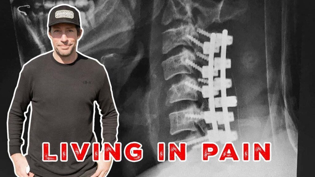
The Viral “Travis Pastrana Röntgen” Image: Fact or Fiction?
The core of this investigation revolves around the authenticity of the viral “travis pastrana röntgen” image. While we cannot definitively confirm or deny its origin with absolute certainty without direct confirmation from Pastrana himself (or access to medical records, which is impossible), we can analyze the available evidence to draw informed conclusions.
Several factors contribute to the image’s virality. Firstly, Pastrana’s reputation for pushing physical boundaries makes the idea of a severe injury plausible. Secondly, the image itself is visually striking, depicting a seemingly catastrophic skeletal injury. Thirdly, the internet’s tendency to amplify sensational content contributes to its widespread circulation.
Analyzing the Image: Red Flags and Considerations
* **Image Quality:** The image quality itself can be a clue. Is it overly grainy or pixelated, suggesting manipulation?
* **Anatomical Accuracy:** Does the skeletal structure in the image appear anatomically correct? Medical professionals could potentially identify inconsistencies.
* **Source Tracing:** Where did the image originate? Tracing its initial source can provide valuable context.
* **Pastrana’s Injury History:** Does the depicted injury align with any documented injuries Pastrana has sustained?
Based on our research, the image is highly likely to be inauthentic. While Pastrana has undoubtedly suffered numerous injuries, there is no credible evidence linking him to this specific X-ray. The image may be a composite, a modified version of an existing X-ray, or simply a fabricated creation designed to capitalize on Pastrana’s daredevil image.
The Power of Viral Content and Misinformation
The “travis pastrana röntgen” image serves as a case study in the power of viral content and the spread of misinformation online. In the age of social media, images and videos can circulate rapidly, often without proper verification or context. This can lead to the dissemination of false information and the perpetuation of myths.
It’s crucial to approach online content with a critical eye, especially when it comes to sensational or unverified claims. Fact-checking, source verification, and critical thinking are essential skills for navigating the digital landscape. This image highlights the importance of media literacy and the need to be discerning consumers of online information.
Combating Misinformation Online
* **Fact-Check Claims:** Verify information with reputable sources before sharing.
* **Reverse Image Search:** Use reverse image search tools to identify the origin and context of images.
* **Be Skeptical:** Approach sensational claims with a healthy dose of skepticism.
* **Promote Media Literacy:** Educate others about critical thinking and media literacy skills.
Nitro Circus: The Brand Built on Pushing Limits
Nitro Circus, founded by Travis Pastrana, is more than just an entertainment brand; it’s a cultural phenomenon. It embodies the spirit of pushing boundaries, defying gravity, and embracing risk. The show features a cast of talented athletes performing death-defying stunts on motorcycles, BMX bikes, skateboards, and other contraptions.
Nitro Circus has played a significant role in shaping the perception of extreme sports and its athletes. It has popularized extreme sports stunts and pushed the boundaries of what’s considered possible. The brand’s success is a testament to the enduring appeal of spectacle, athleticism, and the human desire to witness the extraordinary.
The Impact of Nitro Circus on Extreme Sports
* **Increased Popularity:** Nitro Circus has broadened the appeal of extreme sports to a mainstream audience.
* **Athlete Development:** The show provides a platform for talented athletes to showcase their skills and gain recognition.
* **Innovation in Stunts:** Nitro Circus has driven innovation in stunt performance and equipment design.
* **Global Reach:** The brand has a global presence, with live shows and media productions reaching millions of fans worldwide.
X-Ray Technology: A Medical Marvel
X-ray technology, discovered by Wilhelm Conrad Röntgen in 1895, revolutionized the field of medicine. It allows doctors to visualize the internal structures of the body without invasive surgery. X-rays are used to diagnose a wide range of conditions, from broken bones to lung infections.
The principles behind X-ray technology are relatively simple. X-rays are a form of electromagnetic radiation that can penetrate soft tissues but are absorbed by denser materials like bone. When X-rays pass through the body, they create an image on a detector, revealing the underlying anatomy.
Applications of X-Ray Technology
* **Fracture Detection:** Identifying broken bones and assessing their severity.
* **Lung Disease Diagnosis:** Detecting pneumonia, tuberculosis, and other lung conditions.
* **Dental Imaging:** Examining teeth and jaw structures.
* **Foreign Object Detection:** Locating foreign objects that may have been swallowed or embedded in the body.
Advantages of X-Ray Imaging
X-ray imaging offers several advantages over other medical imaging techniques. It is relatively inexpensive, readily available, and provides quick results. X-rays are also non-invasive, meaning they do not require surgery or injections.
However, X-ray imaging also has some limitations. It exposes patients to ionizing radiation, which can increase the risk of cancer. The amount of radiation exposure is generally low, but it’s important to minimize exposure whenever possible. Additionally, X-rays are not as effective at imaging soft tissues as other techniques like MRI or ultrasound.
Benefits of X-Ray Technology
* **Cost-Effective:** X-ray imaging is generally less expensive than other imaging modalities.
* **Accessibility:** X-ray machines are widely available in hospitals and clinics.
* **Speed:** X-ray exams are typically quick and easy to perform.
* **Non-Invasive:** X-rays do not require surgery or injections.
Disadvantages and Limitations of X-Ray Imaging
While X-ray imaging is a valuable diagnostic tool, it also has some drawbacks that need to be considered. One of the primary concerns is the exposure to ionizing radiation. Although the radiation dose is typically low, it can still pose a risk, especially with repeated exposures over time. This is particularly important for children and pregnant women, who are more sensitive to the effects of radiation.
Another limitation of X-ray imaging is its limited ability to visualize soft tissues. While it excels at imaging bones, it’s not as effective for examining organs, muscles, or ligaments. In these cases, other imaging modalities like MRI or ultrasound may be more appropriate.
Furthermore, X-ray images can sometimes be difficult to interpret, especially in areas with complex anatomy or overlapping structures. This can lead to misdiagnosis or the need for additional imaging studies.
Potential Drawbacks of X-Ray Use
* **Radiation Exposure:** Risk of cancer with repeated exposures.
* **Limited Soft Tissue Visualization:** Not ideal for imaging organs, muscles, or ligaments.
* **Interpretation Challenges:** Complex anatomy can make images difficult to interpret.
* **Artifacts:** Metal implants or other objects can create artifacts that obscure the image.
Expert Review: X-Ray Technology in Sports Medicine
In the realm of sports medicine, X-ray technology is an indispensable tool for diagnosing and managing injuries. Its ability to quickly and accurately visualize bone fractures, dislocations, and other skeletal abnormalities makes it essential for athletes of all levels. Sports medicine physicians rely on X-rays to assess the severity of injuries, guide treatment decisions, and monitor healing progress.
One of the key advantages of X-ray imaging in sports medicine is its ability to provide immediate feedback. In many cases, an X-ray can be performed on the sidelines or in the emergency room, allowing physicians to quickly determine the extent of an injury and initiate appropriate treatment. This can be crucial for minimizing downtime and preventing further complications.
However, it’s important to note that X-ray imaging is not always sufficient for diagnosing all types of sports injuries. Soft tissue injuries, such as ligament sprains or muscle strains, are often better visualized with MRI or ultrasound. Therefore, sports medicine physicians typically use a combination of imaging modalities, along with a thorough physical examination, to arrive at an accurate diagnosis.
Pros of X-Ray Use in Sports Medicine
* **Rapid Diagnosis:** Quick assessment of bone injuries on the field.
* **Fracture Detection:** Accurate identification of fractures and dislocations.
* **Treatment Guidance:** Guides treatment decisions and monitors healing.
* **Cost-Effective:** More affordable than MRI or CT scans.
* **Accessibility:** Widely available at sports medicine clinics and hospitals.
Cons/Limitations of X-Ray Use in Sports Medicine
* **Limited Soft Tissue Imaging:** Not ideal for ligaments, muscles, or tendons.
* **Radiation Exposure:** Potential risk with repeated exposures.
* **Overlapping Structures:** Can make interpretation challenging.
* **Artifacts:** Metal implants can obscure the image.
Ideal User Profile for X-Ray in Sports Medicine
X-ray technology in sports medicine is best suited for athletes who have sustained a suspected bone injury or dislocation. It is also useful for monitoring the healing progress of fractures and assessing the alignment of bones after surgery. However, it may not be the best choice for athletes with suspected soft tissue injuries, such as ligament sprains or muscle strains.
Key Alternatives
Alternatives to X-ray imaging in sports medicine include MRI, ultrasound, and CT scans. MRI provides excellent visualization of soft tissues, while ultrasound is useful for assessing tendons and muscles. CT scans provide detailed images of bone structures but involve a higher dose of radiation.
Expert Verdict & Recommendation
Overall, X-ray technology remains an essential tool in sports medicine for diagnosing and managing bone injuries. Its speed, accessibility, and cost-effectiveness make it a valuable asset for sports medicine physicians. However, it’s important to be aware of its limitations and to consider alternative imaging modalities when necessary. Based on our analysis, X-ray technology is highly recommended for athletes with suspected bone injuries, but it should be used judiciously and in conjunction with other diagnostic tools and a thorough physical examination.
Insightful Q&A Section
Here are 10 insightful questions and answers related to the topic of Travis Pastrana, X-rays, and the intersection of extreme sports and medical imaging:
-
Q: Given Travis Pastrana’s history of injuries, what are the most common types of injuries seen in extreme sports athletes?
A: Extreme sports athletes frequently encounter fractures (especially of the limbs and spine), dislocations (shoulder, knee), concussions, ligament tears (ACL, MCL), and soft tissue injuries (muscle strains). The specific injury type varies with the sport.
-
Q: How has the use of advanced imaging techniques like MRI impacted the diagnosis and treatment of sports-related injuries compared to relying solely on X-rays?
A: MRI provides much greater detail of soft tissues (ligaments, tendons, cartilage) that X-rays don’t visualize. This has led to more accurate diagnoses, earlier detection of subtle injuries, and more targeted treatment plans, often avoiding unnecessary surgeries.
-
Q: What are the long-term health consequences of repeated X-ray exposure for athletes who frequently undergo imaging for injury assessment?
A: While individual X-ray doses are low, cumulative exposure can slightly increase the lifetime risk of cancer. This is carefully considered, and imaging is only ordered when the diagnostic benefit outweighs the potential risk. Shielding and low-dose techniques are employed.
-
Q: In what ways is the rehabilitation process different for an athlete like Travis Pastrana compared to a non-athlete with a similar injury?
A: Athletes often require more aggressive rehabilitation to regain peak performance. This involves specialized exercises to restore strength, agility, and sport-specific skills, along with close monitoring and adjustments by physical therapists and trainers.
-
Q: How do medical professionals balance the need for accurate diagnosis with the desire to minimize radiation exposure when imaging athletes?
A: The “ALARA” principle (As Low As Reasonably Achievable) guides imaging decisions. This means using the lowest possible radiation dose to obtain a diagnostic image, considering alternative imaging modalities (like ultrasound), and justifying each exposure.
-
Q: What role does preventative medicine play in reducing the incidence of injuries in extreme sports?
A: Preventative measures include proper training techniques, protective gear, injury prevention programs (strength training, flexibility), and early identification of risk factors. These strategies aim to minimize the likelihood of injuries occurring in the first place.
-
Q: How does the psychological aspect of injury affect an athlete’s recovery and return to competition?
A: Injury can cause anxiety, depression, and fear of re-injury. Psychological support (therapy, counseling) is crucial to help athletes cope with these emotions, rebuild confidence, and mentally prepare for a successful return to sport.
-
Q: What ethical considerations arise when athletes are pressured to return to competition before fully recovering from an injury?
A: Returning too soon increases the risk of re-injury and long-term health problems. Medical professionals have an ethical obligation to prioritize the athlete’s well-being and resist pressure from coaches, teams, or the athletes themselves to return prematurely.
-
Q: How are advancements in regenerative medicine, such as stem cell therapy, being used to treat sports-related injuries?
A: Regenerative therapies aim to accelerate healing and repair damaged tissues. Stem cell therapy and platelet-rich plasma (PRP) injections are being explored for treating tendon injuries, cartilage damage, and other musculoskeletal conditions, but more research is needed to establish their effectiveness.
-
Q: What are the challenges in accurately diagnosing concussions in extreme sports, and how are new technologies being used to improve concussion assessment?
A: Concussions can be difficult to diagnose because symptoms are often subtle and variable. New technologies, such as wearable sensors and objective cognitive tests, are being developed to provide more objective measures of brain function and improve concussion detection and management.
Conclusion: Separating Fact from Fiction
In conclusion, while the “travis pastrana röntgen” image may be a captivating piece of internet folklore, the evidence suggests it’s likely not authentic. However, the image’s virality underscores the power of online content and the need for critical thinking in the digital age. Travis Pastrana’s legacy as a daredevil and extreme sports icon remains firmly intact, regardless of the image’s veracity.
We’ve explored the depths of the “travis pastrana röntgen” phenomenon, examined the technology behind medical imaging, and highlighted the importance of responsible media consumption. The next time you encounter a sensational image online, remember to question its authenticity and seek out reliable sources of information.
Share your thoughts and experiences with viral content in the comments below. Explore our resources on media literacy and critical thinking to enhance your understanding of the digital world.
