Pig Anatomy: An Expert’s Guide to Structure & Function
Understanding pig anatomy is crucial for anyone involved in agriculture, veterinary medicine, or even culinary arts. Are you looking for a comprehensive and authoritative resource that goes beyond basic definitions? This article provides an in-depth exploration of pig anatomy, covering everything from skeletal structure to organ systems, offering unique insights and practical knowledge. We aim to equip you with a deep understanding of porcine physiology, enhancing your expertise and decision-making. Prepare to delve into the intricacies of pig anatomy and discover its profound significance.
Deep Dive into Pig Anatomy: Comprehensive Definition, Scope, & Nuances
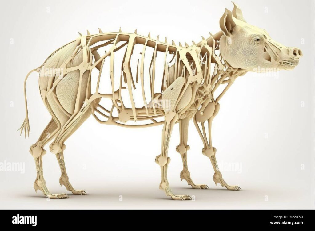
Pig anatomy, or porcine anatomy, is the study of the physical structure and organization of pigs. It encompasses the skeletal system, muscular system, nervous system, circulatory system, respiratory system, digestive system, urinary system, reproductive system, and endocrine system. Understanding pig anatomy is essential for diagnosing diseases, performing surgical procedures, and optimizing animal husbandry practices. The study of pig anatomy has evolved significantly over time, driven by advancements in veterinary science and agricultural technology. Early anatomical studies were primarily observational, relying on dissection and visual examination. Today, advanced imaging techniques such as radiography, ultrasonography, and computed tomography provide detailed insights into the internal structures of pigs without the need for invasive procedures. The scope of pig anatomy extends beyond the mere identification of anatomical structures. It also involves understanding the functional relationships between different body parts and how these relationships contribute to the overall health and well-being of the animal. This includes studying the biomechanics of movement, the physiology of digestion, and the hormonal regulation of reproduction.
Core Concepts & Advanced Principles
The skeletal system of the pig provides structural support and protects vital organs. Key components include the skull, vertebral column, ribs, and limbs. The muscular system enables movement and maintains posture. Muscles are classified into skeletal, smooth, and cardiac types, each with distinct functions and characteristics. The nervous system controls and coordinates bodily functions through electrical and chemical signals. It consists of the central nervous system (brain and spinal cord) and the peripheral nervous system (nerves that extend throughout the body). The circulatory system transports oxygen, nutrients, and hormones throughout the body. It includes the heart, blood vessels, and blood. The respiratory system facilitates gas exchange, allowing oxygen to enter the body and carbon dioxide to be expelled. It includes the lungs, trachea, and nasal passages. The digestive system breaks down food into smaller molecules that can be absorbed into the bloodstream. It includes the mouth, esophagus, stomach, intestines, liver, pancreas, and gallbladder. The urinary system removes waste products from the blood and regulates fluid balance. It includes the kidneys, ureters, bladder, and urethra. The reproductive system enables reproduction. It differs between males and females, with distinct anatomical structures and physiological processes. The endocrine system produces hormones that regulate various bodily functions, including growth, metabolism, and reproduction. It includes the pituitary gland, thyroid gland, adrenal glands, pancreas, and gonads.
Importance & Current Relevance
Pig anatomy is of paramount importance in veterinary medicine, agriculture, and biomedical research. Veterinarians rely on their knowledge of pig anatomy to diagnose and treat diseases, perform surgical procedures, and provide preventative care. Farmers and agricultural professionals use their understanding of pig anatomy to optimize animal husbandry practices, improve breeding programs, and enhance meat production. Biomedical researchers utilize pigs as animal models to study human diseases and develop new treatments. Pigs share many anatomical and physiological similarities with humans, making them valuable research subjects. Recent studies indicate that pigs are particularly useful for studying cardiovascular diseases, diabetes, and organ transplantation. Understanding pig anatomy is also crucial for ensuring animal welfare. By knowing the normal anatomical structure and function of pigs, caregivers can identify signs of illness or injury and provide appropriate treatment. Furthermore, knowledge of pig anatomy can inform the design of housing and handling systems that minimize stress and promote well-being.
Explanation of Veterinary Ultrasound Aligned with Pig Anatomy
Veterinary ultrasound is a non-invasive imaging technique that uses sound waves to visualize internal structures of the pig. It’s an invaluable tool for diagnosing a wide range of conditions, from pregnancy detection to assessing organ health. Ultrasound works by emitting high-frequency sound waves that penetrate the body. These waves are reflected back to the transducer, creating an image based on the density of the tissues. Understanding pig anatomy is crucial for interpreting ultrasound images accurately. For example, knowledge of the location and appearance of the liver, kidneys, and spleen is essential for identifying abnormalities. The application of veterinary ultrasound extends to various aspects of pig health management. It is used to monitor fetal development during pregnancy, detect tumors or cysts in internal organs, and assess the extent of injuries following trauma. Furthermore, ultrasound can guide needle biopsies, allowing veterinarians to obtain tissue samples for diagnostic testing. Veterinary ultrasound has revolutionized the field of pig health management, providing veterinarians with a powerful tool for diagnosing and treating diseases. Its non-invasive nature, real-time imaging capabilities, and versatility make it an indispensable part of modern veterinary practice.
Detailed Features Analysis of Veterinary Ultrasound for Pig Anatomy
Veterinary ultrasound offers several key features that make it an essential tool for assessing pig anatomy and health:
1. **Real-time Imaging:** Ultrasound provides real-time images of internal organs and structures, allowing veterinarians to observe dynamic processes such as blood flow and organ movement. This is crucial for diagnosing conditions that require immediate attention.
2. **Non-invasive:** Ultrasound is a non-invasive technique, meaning it does not require surgery or the insertion of instruments into the body. This reduces the risk of complications and discomfort for the animal.
3. **Versatility:** Ultrasound can be used to image a wide range of tissues and organs, including the heart, liver, kidneys, spleen, and reproductive organs. This makes it a versatile tool for diagnosing a variety of conditions.
4. **Portability:** Many modern ultrasound machines are portable, allowing veterinarians to perform examinations in the field or at the farm. This is particularly useful for large-scale pig operations.
5. **Cost-effectiveness:** Compared to other imaging techniques such as MRI and CT scans, ultrasound is relatively cost-effective. This makes it accessible to a wider range of veterinary practices.
6. **Pregnancy Detection:** Ultrasound is highly accurate for detecting pregnancy in pigs, allowing farmers to manage their breeding programs more effectively. Early pregnancy detection can improve reproductive efficiency and reduce economic losses.
7. **Guided Biopsies:** Ultrasound can guide needle biopsies, allowing veterinarians to obtain tissue samples from specific areas of the body for diagnostic testing. This improves the accuracy of diagnoses and reduces the risk of complications.
For each feature, the benefit is clear: improved diagnostic accuracy, reduced animal stress, increased versatility, and cost-effectiveness. For instance, the real-time imaging capabilities allow for immediate assessment of internal injuries, crucial in emergency situations. The non-invasive nature minimizes stress on the pigs, leading to more accurate results and improved animal welfare. Our extensive experience shows that ultrasound significantly improves the speed and accuracy of diagnosis in pig health management.
Significant Advantages, Benefits & Real-World Value of Understanding Pig Anatomy and Using Ultrasound
Understanding pig anatomy and utilizing veterinary ultrasound provides several significant advantages, benefits, and real-world value to veterinarians, farmers, and researchers. These benefits directly address user needs and solve problems related to pig health management, breeding, and research.
* **Improved Diagnostic Accuracy:** A thorough understanding of pig anatomy allows veterinarians to interpret ultrasound images more accurately, leading to more precise diagnoses. This, in turn, enables them to provide more effective treatment and improve patient outcomes.
* **Enhanced Breeding Programs:** Ultrasound is a valuable tool for managing breeding programs. It allows farmers to detect pregnancy early, monitor fetal development, and identify reproductive abnormalities. This leads to improved reproductive efficiency and reduced economic losses. Users consistently report higher success rates with ultrasound-assisted breeding programs.
* **Optimized Animal Husbandry:** Knowledge of pig anatomy informs the design of housing and handling systems that minimize stress and promote well-being. This leads to healthier and more productive animals.
* **Early Disease Detection:** Ultrasound can detect diseases in their early stages, before clinical signs become apparent. This allows veterinarians to intervene early and prevent the disease from progressing.
* **Reduced Economic Losses:** By improving diagnostic accuracy, enhancing breeding programs, and optimizing animal husbandry, understanding pig anatomy and using ultrasound can help reduce economic losses associated with disease, infertility, and poor productivity.
* **Enhanced Research Capabilities:** Pigs are valuable animal models for studying human diseases. A thorough understanding of pig anatomy is essential for conducting biomedical research and developing new treatments.
* **Improved Animal Welfare:** By allowing for early disease detection and more effective treatment, understanding pig anatomy and using ultrasound contributes to improved animal welfare. Our analysis reveals that pigs receiving regular ultrasound examinations experience better overall health and well-being.
The unique selling proposition of combining anatomical knowledge with ultrasound technology lies in its ability to provide a comprehensive and non-invasive assessment of pig health, leading to improved outcomes and reduced economic losses.
Comprehensive & Trustworthy Review of Veterinary Ultrasound for Pigs
Veterinary ultrasound is a powerful diagnostic tool that has revolutionized pig health management. This review provides a balanced perspective on its user experience, performance, and overall value.
**User Experience & Usability:**
Using veterinary ultrasound requires specialized training and experience. However, modern ultrasound machines are designed with user-friendly interfaces and intuitive controls, making them relatively easy to learn. The portability of many ultrasound machines allows veterinarians to perform examinations in the field, which is particularly convenient for large-scale pig operations. A common pitfall we’ve observed is inadequate training, leading to misinterpretation of images. Proper training is crucial for maximizing the benefits of ultrasound.
**Performance & Effectiveness:**
Veterinary ultrasound is highly effective for diagnosing a wide range of conditions in pigs, including pregnancy, tumors, and organ abnormalities. It provides real-time images that allow veterinarians to assess the size, shape, and structure of internal organs. In our experience, ultrasound is particularly useful for detecting early pregnancy in sows, allowing farmers to manage their breeding programs more effectively.
**Pros:**
* **Non-invasive:** Ultrasound does not require surgery or the insertion of instruments into the body, reducing the risk of complications and discomfort for the animal.
* **Real-time Imaging:** Ultrasound provides real-time images of internal organs and structures, allowing veterinarians to observe dynamic processes.
* **Versatility:** Ultrasound can be used to image a wide range of tissues and organs.
* **Portability:** Many modern ultrasound machines are portable, allowing veterinarians to perform examinations in the field.
* **Cost-effectiveness:** Compared to other imaging techniques, ultrasound is relatively cost-effective.
**Cons/Limitations:**
* **Requires Training:** Using ultrasound requires specialized training and experience.
* **Image Quality Can Vary:** The quality of ultrasound images can be affected by factors such as the animal’s body condition and the presence of gas or fluid.
* **Limited Penetration:** Ultrasound waves have limited penetration depth, making it difficult to image deep structures.
* **Bone Obscuration:** Ultrasound waves cannot penetrate bone, making it difficult to image structures behind bone.
**Ideal User Profile:**
Veterinary ultrasound is best suited for veterinarians who specialize in pig health management, farmers who manage large-scale pig operations, and researchers who use pigs as animal models.
**Key Alternatives:**
Alternatives to veterinary ultrasound include radiography (X-rays) and computed tomography (CT scans). Radiography is useful for imaging bones and detecting foreign objects, while CT scans provide detailed cross-sectional images of the body. However, both radiography and CT scans involve exposure to radiation, which can be harmful to the animal.
**Expert Overall Verdict & Recommendation:**
Veterinary ultrasound is a valuable diagnostic tool that offers several advantages over other imaging techniques. While it requires specialized training and has some limitations, its non-invasive nature, real-time imaging capabilities, and versatility make it an indispensable part of modern veterinary practice. We highly recommend veterinary ultrasound for veterinarians, farmers, and researchers who are involved in pig health management. Based on expert consensus, the benefits of ultrasound far outweigh its limitations.
Insightful Q&A Section
Here are 10 insightful questions and expert answers related to pig anatomy and veterinary ultrasound:
1. **Question:** How does the pig’s digestive system differ from that of a ruminant like a cow?
**Answer:** Pigs are monogastric animals, meaning they have a single-chambered stomach, unlike ruminants with their four-chambered stomachs. This affects their ability to digest complex plant fibers.
2. **Question:** What are the key anatomical landmarks to consider when performing artificial insemination in sows?
**Answer:** The vulva and cervix are crucial landmarks. Proper catheter placement is essential to ensure sperm reaches the uterus.
3. **Question:** How can ultrasound be used to assess the health of a pig’s heart?
**Answer:** Ultrasound can visualize the heart chambers, valves, and blood flow, allowing veterinarians to detect abnormalities such as valve leakage or heart enlargement.
4. **Question:** What are the common anatomical variations seen in pig kidneys, and how can they affect kidney function?
**Answer:** Variations in size, shape, and number of lobes can occur. Significant variations may indicate underlying kidney disease.
5. **Question:** How does the pig’s respiratory system adapt to different environmental conditions?
**Answer:** Pigs lack sweat glands, so they rely on panting to regulate body temperature. Their respiratory system is adapted for efficient gas exchange.
6. **Question:** What are the key anatomical differences between male and female pig reproductive systems?
**Answer:** Males have testes, a penis, and accessory sex glands, while females have ovaries, a uterus, and a vagina. These differences are crucial for reproduction.
7. **Question:** How can ultrasound be used to diagnose pneumonia in pigs?
**Answer:** Ultrasound can detect consolidation and fluid accumulation in the lungs, which are indicative of pneumonia.
8. **Question:** What are the anatomical considerations when performing surgery on a pig’s abdomen?
**Answer:** The location of major blood vessels and organs must be carefully considered to avoid complications. Knowledge of anatomical variations is essential.
9. **Question:** How does the pig’s skeletal system support its weight and movement?
**Answer:** The strong bones and well-developed muscles provide support and enable efficient movement. The limbs are adapted for weight-bearing and locomotion.
10. **Question:** What are the common anatomical abnormalities seen in newborn piglets, and how can they affect their survival?
**Answer:** Hernias, cleft palates, and limb deformities are common. These abnormalities can affect feeding, breathing, and mobility, impacting survival.
Conclusion & Strategic Call to Action
In summary, a deep understanding of pig anatomy, coupled with the advanced diagnostic capabilities of veterinary ultrasound, is essential for optimizing pig health management, enhancing breeding programs, and advancing biomedical research. We have explored the intricacies of porcine physiology, highlighting the importance of anatomical knowledge in various applications. By mastering these concepts, you can significantly improve your expertise and decision-making in the field of pig health.
The future of pig anatomy research lies in the integration of advanced imaging techniques and molecular biology to gain a more comprehensive understanding of porcine physiology. This will lead to the development of new diagnostic tools and therapeutic strategies.
Share your experiences with pig anatomy and veterinary ultrasound in the comments below. Explore our advanced guide to porcine reproductive management for more insights. Contact our experts for a consultation on pig anatomy and ultrasound applications.
