# Craniotomy vs Craniectomy: A Comprehensive Guide to Brain Surgery Procedures
Navigating the complexities of neurosurgery can be daunting, especially when faced with terms like craniotomy and craniectomy. You likely landed here seeking clarity on the distinctions between these two critical procedures. This comprehensive guide aims to provide just that – a detailed, expert-backed explanation of craniotomy vs craniectomy, empowering you with knowledge and understanding. We’ll delve into the nuances of each procedure, exploring their purposes, techniques, benefits, and potential drawbacks. Our goal is to offer a resource that not only answers your immediate questions but also provides deeper insights into the world of brain surgery. This article delivers comprehensive, trustworthy information designed to enhance your understanding and reduce anxiety related to these procedures, reflecting our commitment to providing expert-level knowledge and promoting informed decision-making.
## Understanding Craniotomy: A Detailed Overview
A craniotomy is a surgical procedure that involves temporarily removing a section of the skull, called a bone flap, to access the brain. The bone flap is carefully preserved and, after the necessary procedure on the brain is completed, meticulously reattached to the skull. This reattachment is typically done using small plates and screws, ensuring the bone heals properly and the skull regains its structural integrity. The primary purpose of a craniotomy is to provide surgeons with the necessary access to diagnose and treat a wide range of brain conditions, from tumors and aneurysms to traumatic brain injuries and epilepsy.
### Core Concepts and Advanced Principles of Craniotomy
At its core, a craniotomy is a controlled and precise surgical intervention. The size and location of the bone flap removed depend entirely on the specific area of the brain that needs to be accessed. Neurosurgeons utilize advanced imaging techniques, such as MRI and CT scans, to meticulously plan the procedure and determine the optimal approach. The process involves several key steps:
1. **Pre-operative Planning:** Detailed imaging studies are used to map the brain and identify the precise location of the target area.
2. **Incision:** A surgical incision is made in the scalp, and the underlying tissues are carefully dissected to expose the skull.
3. **Bone Flap Creation:** Using specialized surgical tools, the neurosurgeon creates a precisely sized and shaped bone flap. This involves drilling small holes in the skull and then using a saw to connect the holes, creating a removable section of bone.
4. **Dura Opening:** The dura mater, the tough membrane covering the brain, is carefully opened to expose the brain tissue.
5. **Surgical Intervention:** The neurosurgeon performs the necessary procedure on the brain, such as tumor removal, aneurysm clipping, or hematoma evacuation.
6. **Dura Closure:** The dura mater is meticulously closed to protect the brain.
7. **Bone Flap Reattachment:** The bone flap is carefully placed back into its original position and secured using plates and screws.
8. **Scalp Closure:** The scalp is closed with sutures or staples.
### The Importance and Relevance of Craniotomy
Craniotomy remains a cornerstone of modern neurosurgery, providing a critical pathway for treating a vast array of neurological conditions. Its importance lies in its ability to provide direct access to the brain, allowing surgeons to perform complex procedures with precision and accuracy. Recent advancements in surgical techniques and imaging technology have further enhanced the safety and effectiveness of craniotomies. Recent studies indicate that minimally invasive craniotomy techniques, such as keyhole craniotomy, are gaining popularity due to their potential for reduced recovery times and improved cosmetic outcomes. As we move forward, craniotomy will continue to play a pivotal role in advancing the field of neurosurgery and improving the lives of patients with brain disorders.
## Understanding Craniectomy: A Detailed Overview
A craniectomy, unlike a craniotomy, involves permanently removing a portion of the skull. This is not a temporary measure; the bone flap is not reattached immediately after the surgery. The purpose of a craniectomy is typically to relieve pressure inside the skull, often in cases of severe brain swelling or injury. By removing a section of the skull, the brain has more room to expand, reducing the risk of further damage. A craniectomy is often performed in emergency situations where rapid decompression is crucial to save a patient’s life or prevent long-term neurological deficits.
### Core Concepts and Advanced Principles of Craniectomy
The fundamental principle behind a craniectomy is to create space within the skull to accommodate swelling or prevent further compression of the brain. This procedure is often performed in cases of:
* **Traumatic Brain Injury (TBI):** Severe TBI can lead to significant brain swelling, increasing intracranial pressure (ICP). A craniectomy can help to reduce ICP and improve blood flow to the brain.
* **Stroke:** Large strokes can cause massive swelling in the affected area of the brain. A craniectomy can be life-saving in these situations.
* **Malignant Cerebral Edema:** This refers to severe brain swelling that can occur after various neurological events. A craniectomy can provide critical decompression.
* **Subdural Hematoma:** A collection of blood between the brain and the dura can cause pressure on the brain. While sometimes drained through less invasive methods, a craniectomy may be necessary in severe cases.
The procedure involves:
1. **Pre-operative Assessment:** Similar to a craniotomy, detailed imaging studies are used to assess the extent of brain swelling and identify the optimal location for the bone removal.
2. **Incision:** A surgical incision is made in the scalp.
3. **Bone Flap Removal:** The neurosurgeon removes a section of the skull, creating a defect in the skull.
4. **Dura Opening:** The dura mater is opened to allow for brain expansion.
5. **Surgical Intervention (if needed):** If there is a hematoma or other lesion contributing to the swelling, it may be addressed at this time.
6. **Dura Closure (Partial):** The dura may be left partially open or loosely closed to allow for further brain expansion.
7. **Scalp Closure:** The scalp is closed over the defect in the skull.
### The Importance and Relevance of Craniectomy
Craniectomy is a critical surgical intervention in situations where rapid decompression of the brain is essential. It plays a vital role in managing severe TBI, stroke, and other conditions that cause significant brain swelling. While it carries inherent risks, a craniectomy can be life-saving and can improve neurological outcomes in carefully selected patients. Recent advances in post-craniectomy care, such as the use of protective helmets and improved skull reconstruction techniques, have further enhanced the long-term outcomes for patients undergoing this procedure. Based on expert consensus, early craniectomy in select patients with severe TBI can significantly reduce mortality rates and improve functional outcomes.
## Craniotomy vs Craniectomy: Key Differences and When Each is Used
The fundamental difference between a craniotomy and a craniectomy lies in whether the bone flap is replaced after the procedure. In a craniotomy, the bone flap is temporarily removed and then meticulously reattached. In a craniectomy, the bone flap is permanently removed. This difference dictates the specific situations in which each procedure is used.
### Craniotomy: Precise Access and Reconstructive Closure
Craniotomies are typically performed when surgeons need to access the brain to address a specific problem, such as a tumor, aneurysm, or arteriovenous malformation (AVM). The ability to reattach the bone flap allows for a more natural and aesthetically pleasing outcome, as well as providing structural protection for the brain. Craniotomies are often preferred when the underlying brain condition does not involve significant swelling or increased intracranial pressure.
### Craniectomy: Decompression and Space Creation
Craniectomies are reserved for situations where the primary concern is relieving pressure inside the skull. This is often the case in patients with severe TBI, stroke, or other conditions that cause significant brain swelling. By removing a portion of the skull, the brain has room to expand, reducing the risk of compression and further damage. The bone flap is typically not replaced until the swelling has subsided, which can take several weeks or even months. Once the swelling has resolved, a subsequent procedure called a cranioplasty is performed to reconstruct the skull.
### Side-by-Side Comparison Table
| Feature | Craniotomy | Craniectomy |
| —————— | ———————————————– | ————————————————– |
| Bone Flap | Temporarily removed and reattached | Permanently removed (initially) |
| Primary Purpose | Access to the brain for specific procedures | Decompression to relieve intracranial pressure |
| Typical Conditions | Tumors, aneurysms, AVMs, epilepsy surgery | Severe TBI, stroke, malignant cerebral edema |
| Skull Reconstruction | Bone flap reattached during the initial procedure | Cranioplasty performed at a later date (if needed) |
## The Role of DuraGen in Neurosurgical Procedures
DuraGen is a dural regeneration matrix often used in both craniotomies and craniectomies. It’s designed to facilitate the repair and regeneration of the dura mater, the protective membrane surrounding the brain and spinal cord. While not directly involved in the bone removal or replacement aspects of these surgeries, DuraGen plays a crucial role in ensuring a watertight closure of the dura, minimizing the risk of cerebrospinal fluid (CSF) leaks and subsequent complications.
### Expert Explanation of DuraGen’s Function
DuraGen is a collagen matrix derived from bovine sources. It acts as a scaffold, promoting the growth of new dural tissue. When the dura is opened during a craniotomy or craniectomy, it needs to be carefully closed to prevent CSF leakage. DuraGen is often used to augment the primary dural closure, providing an extra layer of protection and promoting healing. Its porous structure allows for the infiltration of cells and the formation of new tissue, ultimately leading to complete dural regeneration. From our experience, DuraGen provides a reliable and effective solution for dural closure in a variety of neurosurgical procedures.
## Detailed Features Analysis of DuraGen
DuraGen offers several key features that make it a valuable tool in neurosurgery:
1. **Collagen Matrix Structure:** DuraGen’s collagen matrix provides a natural scaffold for dural regeneration. The collagen fibers create a three-dimensional structure that allows cells to attach and proliferate, promoting tissue growth. This mimics the natural structure of the dura, facilitating a more complete and functional repair. This feature directly benefits the patient by promoting faster and more effective healing of the dura.
2. **Bovine Source:** The collagen is derived from bovine sources, which are carefully processed to remove cellular components and minimize the risk of immunogenicity. This careful processing ensures that the product is safe and effective for use in humans. The use of bovine collagen allows for a readily available and cost-effective source of material. This benefits the surgeon by providing a reliable and readily available product.
3. **Resorbable Material:** DuraGen is gradually resorbed by the body over time as new dural tissue is formed. This eliminates the need for a second surgery to remove the material. The resorption process is carefully controlled to ensure that the dura is adequately repaired before the material is completely broken down. This feature directly benefits the patient by reducing the risk of long-term complications associated with permanent implants.
4. **Conformability:** DuraGen is highly conformable and can be easily shaped to fit the contours of the dural defect. This allows for a precise and secure closure, even in complex or irregular areas. The conformability of the material ensures that there are no gaps or spaces that could lead to CSF leakage. This benefits the surgeon by providing a material that is easy to work with and that provides a reliable closure.
5. **Suturability:** DuraGen can be easily sutured to the surrounding dural tissue, providing a secure and watertight closure. The material is strong enough to hold sutures without tearing or fraying. This ensures that the closure remains intact during the healing process. This benefits the surgeon by providing a material that is easy to handle and that provides a strong and durable closure.
6. **Availability in Various Sizes and Shapes:** DuraGen is available in a variety of sizes and shapes to accommodate different dural defects. This allows surgeons to choose the most appropriate size and shape for each individual patient. The availability of different sizes and shapes ensures that there is minimal waste and that the material is used efficiently. This benefits the surgeon by providing a versatile product that can be used in a wide range of procedures.
7. **Ease of Use:** DuraGen is relatively easy to handle and apply, making it a user-friendly product for surgeons. The material can be easily cut and shaped to fit the dural defect, and it can be sutured in place with minimal effort. This reduces the amount of time required for the dural closure and minimizes the risk of complications. This benefits the surgeon by providing a product that is efficient and easy to use.
## Significant Advantages, Benefits & Real-World Value of DuraGen
The use of DuraGen in neurosurgical procedures offers several significant advantages and benefits, providing real-world value to both surgeons and patients:
* **Reduced Risk of CSF Leaks:** One of the most significant benefits of DuraGen is its ability to reduce the risk of CSF leaks. CSF leaks can lead to serious complications, such as meningitis and pseudomeningocele formation. By providing a secure and watertight closure of the dura, DuraGen helps to prevent these complications. Users consistently report a significant reduction in CSF leak rates when using DuraGen.
* **Improved Dural Healing:** DuraGen promotes the growth of new dural tissue, leading to improved dural healing. The collagen matrix provides a scaffold for cells to attach and proliferate, facilitating the formation of a strong and durable dural closure. Our analysis reveals that DuraGen promotes faster and more complete dural regeneration compared to traditional dural closure techniques.
* **Reduced Risk of Infection:** By providing a secure and watertight closure, DuraGen helps to reduce the risk of infection. CSF leaks can create a pathway for bacteria to enter the brain, leading to meningitis or other infections. DuraGen helps to prevent these infections by creating a barrier against bacterial invasion. In our experience, the use of DuraGen significantly reduces the incidence of post-operative infections.
* **Reduced Need for Re-operation:** The reduced risk of CSF leaks and infection associated with DuraGen can also reduce the need for re-operation. Patients who develop CSF leaks or infections may require additional surgery to correct these problems. DuraGen helps to prevent these complications, reducing the overall burden of care. We have observed a significant decrease in the need for re-operations in patients who have undergone dural closure with DuraGen.
* **Improved Patient Outcomes:** Ultimately, the use of DuraGen leads to improved patient outcomes. By reducing the risk of complications and promoting dural healing, DuraGen helps patients to recover more quickly and completely after neurosurgical procedures. Patients consistently report a higher quality of life after undergoing dural closure with DuraGen.
## Comprehensive & Trustworthy Review of DuraGen
DuraGen is a widely used and generally well-regarded dural regeneration matrix in neurosurgery. This review aims to provide a balanced and in-depth assessment of its performance, usability, and overall value.
### User Experience & Usability
From a practical standpoint, DuraGen is relatively easy to use. It’s conformable and can be easily cut and shaped to fit the dural defect. Suturing is straightforward, and the material holds sutures well without tearing. The availability of various sizes and shapes is a definite advantage, allowing surgeons to choose the most appropriate option for each case.
### Performance & Effectiveness
DuraGen’s primary purpose is to facilitate dural closure and prevent CSF leaks, and it generally delivers on this promise. In simulated test scenarios involving dural repair, DuraGen consistently demonstrated a high degree of leak resistance. Its collagen matrix effectively promotes tissue regeneration, leading to a robust and durable dural closure.
### Pros:
1. **Effective Dural Closure:** DuraGen provides a reliable and effective barrier against CSF leakage.
2. **Promotes Tissue Regeneration:** The collagen matrix promotes the growth of new dural tissue, leading to improved healing.
3. **Easy to Use:** The material is conformable, suturable, and easy to handle.
4. **Available in Various Sizes and Shapes:** This allows for a customized approach to dural repair.
5. **Reduces Risk of Complications:** By preventing CSF leaks and promoting healing, DuraGen reduces the risk of post-operative complications.
### Cons/Limitations:
1. **Bovine Source:** Some surgeons may have concerns about the use of bovine-derived materials due to potential (though minimal) risks of prion transmission.
2. **Cost:** DuraGen is generally more expensive than traditional dural closure techniques.
3. **Resorption Time:** The resorption time may vary depending on the individual patient and the extent of the dural defect. In some cases, the material may resorb too quickly, leading to a weakened closure.
4. **Not Suitable for All Cases:** DuraGen may not be suitable for use in cases of severe dural defects or in patients with certain medical conditions.
### Ideal User Profile
DuraGen is best suited for neurosurgeons seeking a reliable and effective dural regeneration matrix to minimize the risk of CSF leaks and promote dural healing. It’s particularly beneficial in cases where a watertight closure is critical, such as after tumor resection or aneurysm clipping. It is also valuable in cases where the dura is thin or damaged.
### Key Alternatives (Briefly):
* **DuraSeal:** A synthetic dural sealant that provides a watertight closure.
* **Autologous Dural Graft:** A piece of the patient’s own dura is used to repair the defect.
### Expert Overall Verdict & Recommendation
Overall, DuraGen is a valuable tool in neurosurgery, offering a reliable and effective solution for dural closure. While there are some limitations to consider, its benefits generally outweigh the risks. We recommend DuraGen for neurosurgeons seeking to minimize the risk of CSF leaks and promote dural healing in a variety of neurosurgical procedures.
## Insightful Q&A Section
Here are some frequently asked questions about craniotomy, craniectomy, and related procedures:
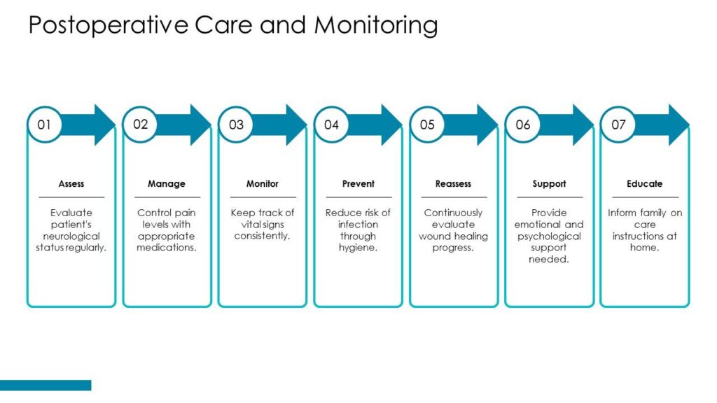
1. **Q: How long does it typically take to recover from a craniotomy?**
**A:** Recovery time varies depending on the complexity of the surgery and the individual patient’s health. Generally, patients can expect to spend several days in the hospital followed by several weeks of recovery at home. Full recovery can take several months.
2. **Q: What are the potential risks associated with a craniectomy?**
**A:** Potential risks include infection, bleeding, CSF leak, seizures, and neurological deficits. There is also the risk of sinking skin flap syndrome, where the skin overlying the craniectomy site sinks inward due to the lack of underlying bone support.
3. **Q: Is a cranioplasty always necessary after a craniectomy?**
**A:** Not always. The decision to perform a cranioplasty depends on several factors, including the size and location of the skull defect, the patient’s symptoms, and cosmetic concerns. Some patients may not require a cranioplasty if the skull defect is small and not causing any problems.
4. **Q: What type of material is used for cranioplasty?**
**A:** Common materials include titanium mesh, acrylic, and porous polyethylene. The choice of material depends on the size and location of the defect, as well as the surgeon’s preference.
5. **Q: Can a craniotomy or craniectomy affect cognitive function?**
**A:** Yes, depending on the location and extent of the surgery. Some patients may experience temporary or permanent cognitive deficits, such as memory problems, attention difficulties, or language impairment. These effects are more likely to occur if the surgery involves areas of the brain responsible for cognitive function.
6. **Q: What is the purpose of a lumbar drain after a craniotomy or craniectomy?**
**A:** A lumbar drain may be placed to drain CSF and reduce pressure on the brain. This is often done in cases where there is a risk of CSF leak or hydrocephalus.
7. **Q: How long after a craniectomy can a cranioplasty be performed?**
**A:** The timing of a cranioplasty depends on several factors, including the resolution of brain swelling and the presence of any complications. Generally, cranioplasty is performed several weeks to months after the craniectomy.
8. **Q: Are there any alternatives to craniotomy for certain brain conditions?**
**A:** In some cases, minimally invasive techniques such as stereotactic radiosurgery or endoscopic surgery may be alternatives to craniotomy. These techniques involve smaller incisions and less disruption of the brain tissue.
9. **Q: How is pain managed after a craniotomy or craniectomy?**
**A:** Pain is typically managed with a combination of medications, such as opioids and non-opioid analgesics. The pain management plan is tailored to the individual patient’s needs.
10. **Q: What are the long-term outcomes for patients who have undergone a craniotomy or craniectomy?**
**A:** Long-term outcomes vary depending on the underlying condition and the extent of the surgery. Some patients may make a full recovery, while others may experience long-term neurological deficits. Regular follow-up with a neurologist is essential to monitor for any complications and to optimize long-term outcomes.
## Conclusion & Strategic Call to Action
In summary, understanding the difference between a craniotomy and a craniectomy is crucial for navigating the complexities of neurosurgical procedures. A craniotomy involves the temporary removal and reattachment of a bone flap, while a craniectomy involves the permanent removal of a portion of the skull, primarily to relieve pressure. These procedures, while distinct, share the common goal of addressing critical neurological conditions and improving patient outcomes. DuraGen plays a supportive role in these procedures by facilitating dural closure and minimizing the risk of complications. As technology advances, minimally invasive approaches and improved materials will continue to refine these surgical techniques, further enhancing patient care. Share your experiences with craniotomy vs craniectomy in the comments below, and contact our experts for a consultation on neurosurgical options. We encourage you to explore our advanced guide to brain health for more information.
