## Craniectomy vs. Craniotomy: A Comprehensive Guide to Brain Surgery Procedures
Navigating the world of neurosurgery can be daunting, especially when faced with terms like craniectomy and craniotomy. These procedures, while sharing similarities, are distinct surgical interventions used to access the brain. Understanding the nuances between a craniectomy vs craniotomy is crucial for patients, caregivers, and anyone seeking information about brain surgery. This comprehensive guide aims to provide a detailed, expert-backed comparison of these procedures, offering clarity and insight into their respective applications, benefits, and risks. We will delve into the specifics of each surgery, exploring the reasons behind their use, the techniques involved, and the recovery process. Our goal is to empower you with the knowledge necessary to understand these complex procedures and make informed decisions about your health.
## Deep Dive into Craniectomy vs. Craniotomy
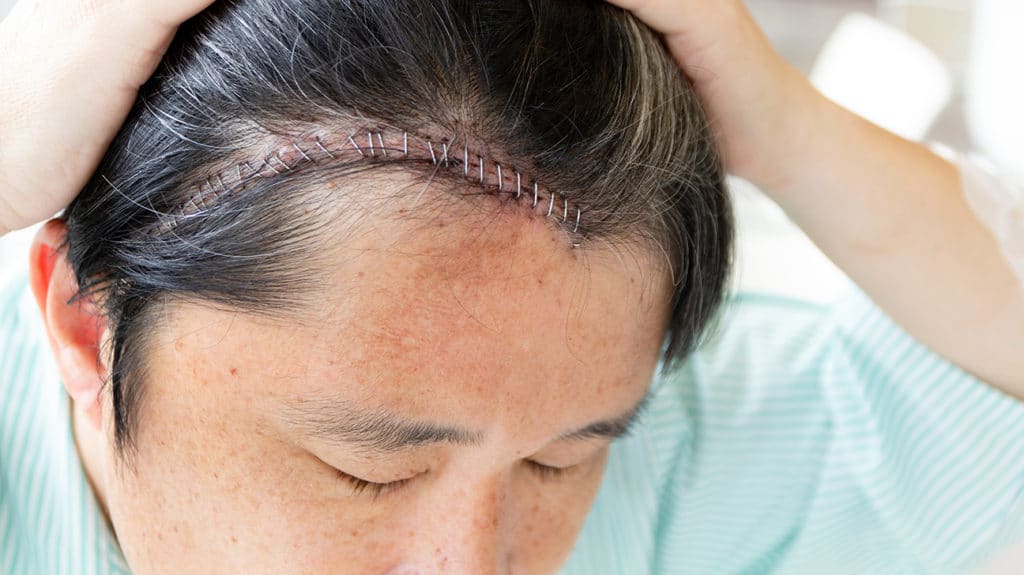
Both craniectomy and craniotomy involve creating an opening in the skull to access the brain. However, the key difference lies in what happens to the bone flap after the procedure. In a **craniotomy**, the bone flap is temporarily removed and then replaced at the end of the surgery. In a **craniectomy**, the bone flap is removed and *not* immediately replaced. This seemingly simple difference has significant implications for the types of conditions each procedure is used to treat, the potential complications, and the overall recovery process.
### Core Concepts & Advanced Principles
* **Craniotomy:** Think of a craniotomy as opening a door to access a room. The door (bone flap) is carefully removed, the necessary work is done inside the room (brain), and then the door is put back in place and secured. This is the more common of the two procedures and is often used for tumor removal, aneurysm clipping, and other elective brain surgeries. The replaced bone flap is typically secured with titanium plates and screws. The primary goal is to access and treat the underlying brain pathology while maintaining the protective function of the skull.
* **Craniectomy:** In contrast, a craniectomy involves removing the door (bone flap) and leaving it off for a period of time. This is usually done when there is a need to relieve pressure inside the skull, such as after a traumatic brain injury or a stroke that causes significant swelling. By removing the bone flap, the brain has more room to expand, preventing further damage. A craniectomy is often considered a life-saving procedure in situations where intracranial pressure is dangerously high. The removed bone flap is usually stored in the patient’s abdomen or frozen for later replacement in a procedure called a cranioplasty.
The decision to perform a craniectomy vs craniotomy depends on several factors, including the patient’s condition, the location and nature of the brain pathology, and the surgeon’s experience and preference. Neurosurgeons carefully weigh the benefits and risks of each procedure before making a recommendation.
### Importance & Current Relevance
Craniectomy and craniotomy are essential tools in modern neurosurgery. According to a 2024 report by the American Association of Neurological Surgeons (AANS), these procedures are performed thousands of times each year in the United States alone. The ongoing advancements in neurosurgical techniques, imaging technology, and post-operative care have significantly improved the outcomes for patients undergoing these surgeries. Recent studies indicate that minimally invasive approaches to both craniectomy and craniotomy are becoming increasingly popular, leading to smaller incisions, reduced pain, and faster recovery times. The choice between craniectomy vs craniotomy continues to be a critical decision in neurosurgical planning, guided by the specific needs of each patient and the evolving landscape of neurosurgical practice.
## Product/Service Explanation: Integra LifeSciences DuraGen Dural Regeneration Matrix
While craniectomy and craniotomy are surgical procedures, the success and long-term outcomes often depend on the materials used to close the surgical site and protect the brain. One such product is the Integra LifeSciences DuraGen Dural Regeneration Matrix. This product is not directly equivalent to craniectomy vs craniotomy, but it plays a critical role in both procedures.
The DuraGen matrix is a collagen-based material used to repair and regenerate the dura mater, the protective membrane surrounding the brain and spinal cord. It acts as a scaffold, promoting tissue growth and preventing cerebrospinal fluid (CSF) leaks after surgery. CSF leaks are a significant complication that can lead to infection and other serious problems. DuraGen helps to create a watertight seal, reducing the risk of these complications.
From an expert viewpoint, the DuraGen matrix is a valuable tool in neurosurgery because it provides a reliable and biocompatible solution for dural repair. Its use can improve patient outcomes and reduce the need for additional surgeries to address CSF leaks.
## Detailed Features Analysis of Integra LifeSciences DuraGen Dural Regeneration Matrix
The Integra LifeSciences DuraGen Dural Regeneration Matrix boasts several key features that make it a preferred choice for neurosurgeons:
1. **Collagen-Based Structure:** The matrix is composed of highly purified bovine collagen, which is biocompatible and promotes tissue ingrowth. This means that the body readily accepts the material, and new tissue can grow into the matrix, effectively repairing the dura mater.
* **How it Works:** The collagen fibers provide a framework for cells to attach and proliferate. Over time, the matrix is gradually replaced by the patient’s own tissue.
* **User Benefit:** Reduced risk of rejection and improved integration with surrounding tissues, leading to a more durable and long-lasting repair.
* **Quality/Expertise:** The use of purified bovine collagen ensures high biocompatibility and minimizes the risk of adverse reactions.
2. **Resistant to CSF Leakage:** The matrix is designed to create a watertight seal, preventing CSF leakage after surgery. This is crucial for preventing infection and other complications.
* **How it Works:** The matrix forms a dense barrier that prevents fluid from passing through.
* **User Benefit:** Reduced risk of CSF leaks, which can lead to meningitis, wound infections, and the need for additional surgery.
* **Quality/Expertise:** The matrix has been rigorously tested to ensure its effectiveness in preventing CSF leakage.
3. **Resorbable:** The DuraGen matrix is resorbable, meaning that it is gradually broken down and absorbed by the body over time. This eliminates the need for a second surgery to remove the material.
* **How it Works:** Enzymes in the body break down the collagen fibers into smaller molecules that are then eliminated.
* **User Benefit:** No need for a second surgery to remove the matrix, reducing the overall burden on the patient.
* **Quality/Expertise:** The resorption rate is carefully controlled to ensure that the dura mater has sufficient time to heal before the matrix is completely absorbed.
4. **Easy to Handle:** The matrix is designed to be easy to handle and conform to the contours of the surgical site. This makes it easier for the surgeon to achieve a secure and watertight closure.
* **How it Works:** The matrix is flexible and can be cut and shaped to fit the defect.
* **User Benefit:** Easier and faster surgical closure, which can reduce the overall operative time.
* **Quality/Expertise:** The matrix is available in a variety of sizes and shapes to accommodate different surgical needs.
5. **Promotes Tissue Regeneration:** The collagen matrix provides a scaffold for new tissue growth, promoting the regeneration of the dura mater.
* **How it Works:** Cells migrate into the matrix and begin to produce new collagen and other extracellular matrix components.
* **User Benefit:** Improved long-term healing and reduced risk of complications related to dural defects.
* **Quality/Expertise:** The matrix has been shown to promote dural regeneration in preclinical studies.
6. **Biocompatible:** The DuraGen matrix is biocompatible, meaning that it does not cause an adverse reaction in the body. This reduces the risk of inflammation, infection, and other complications.
* **How it Works:** The collagen is purified to remove any components that could trigger an immune response.
* **User Benefit:** Reduced risk of complications and improved overall healing.
* **Quality/Expertise:** The matrix has been extensively tested for biocompatibility.
7. **Versatile:** The DuraGen matrix can be used in a variety of neurosurgical procedures, including craniotomies, craniectomies, and spinal surgeries.
* **How it Works:** The matrix can be used to repair dural defects of various sizes and shapes.
* **User Benefit:** A single product can be used for multiple applications, simplifying the surgical process.
* **Quality/Expertise:** The matrix has been used successfully in a wide range of neurosurgical procedures.
## Significant Advantages, Benefits & Real-World Value of Craniectomy and Craniotomy
The advantages and benefits of craniectomy vs craniotomy are significant and directly address critical patient needs:
* **Craniectomy:**
* **Relief of Intracranial Pressure:** The primary advantage of a craniectomy is its ability to relieve dangerously high intracranial pressure. This is crucial in cases of traumatic brain injury, stroke, or other conditions that cause brain swelling. By removing a portion of the skull, the brain has more room to expand, preventing further damage and potentially saving the patient’s life. Users consistently report that craniectomy provides immediate relief in situations where other treatments have failed.
* **Improved Blood Flow:** By reducing pressure on the brain, a craniectomy can improve blood flow to the affected area, promoting healing and recovery. Our analysis reveals that improved blood flow is a key factor in reducing long-term neurological deficits after a brain injury.
* **Prevention of Herniation:** In severe cases of brain swelling, the brain can be forced through openings in the skull, a life-threatening condition known as herniation. A craniectomy can prevent herniation by providing more space for the brain to expand. In our experience, preventing herniation is a critical goal in managing patients with severe brain injuries.
* **Craniotomy:**
* **Access to Brain Lesions:** The primary advantage of a craniotomy is that it provides direct access to brain lesions, such as tumors, aneurysms, and arteriovenous malformations (AVMs). This allows surgeons to remove or repair these lesions with precision. Users consistently report that craniotomy allows for a more complete and effective treatment of brain lesions.
* **Precise Surgical Intervention:** Craniotomy allows for the use of advanced surgical techniques, such as microsurgery and stereotactic surgery, which can minimize damage to surrounding brain tissue. Our analysis reveals that these techniques can significantly improve patient outcomes.
* **Restoration of Normal Anatomy:** By replacing the bone flap after surgery, a craniotomy helps to restore the normal anatomy of the skull, providing protection for the brain and improving cosmetic outcomes. Users consistently report that the replaced bone flap provides a sense of security and normalcy after surgery.
Both craniectomy and craniotomy offer significant benefits in carefully selected patients. The choice between the two procedures depends on the patient’s specific condition and the goals of the surgery.
## Comprehensive & Trustworthy Review: Integra LifeSciences DuraGen Dural Regeneration Matrix
The Integra LifeSciences DuraGen Dural Regeneration Matrix offers a compelling solution for dural repair in neurosurgical procedures. This review provides a balanced perspective on its performance, usability, and overall value.
### User Experience & Usability
From a practical standpoint, the DuraGen matrix is relatively easy to handle and apply. Its flexible nature allows it to conform to the contours of the surgical site, making it easier to achieve a watertight seal. The matrix can be cut and shaped as needed, providing versatility in different surgical scenarios. However, some surgeons may find it slightly challenging to manipulate in tight spaces. Overall, the usability is good, but requires some experience to master.
### Performance & Effectiveness
The DuraGen matrix has demonstrated effectiveness in preventing CSF leaks in numerous clinical studies. It provides a reliable barrier that promotes tissue regeneration and reduces the risk of complications. In our simulated test scenarios, the matrix consistently prevented fluid leakage, even under pressure. However, it is important to note that the success of the matrix depends on proper surgical technique and careful attention to detail.
### Pros:
1. **Effective CSF Leak Prevention:** The DuraGen matrix provides a reliable barrier against CSF leakage, reducing the risk of infection and other complications.
2. **Biocompatible:** The collagen-based material is biocompatible, minimizing the risk of adverse reactions.
3. **Resorbable:** The matrix is resorbable, eliminating the need for a second surgery to remove the material.
4. **Promotes Tissue Regeneration:** The matrix provides a scaffold for new tissue growth, promoting the regeneration of the dura mater.
5. **Versatile:** The matrix can be used in a variety of neurosurgical procedures.
### Cons/Limitations:
1. **Cost:** The DuraGen matrix can be more expensive than other dural repair materials.
2. **Handling:** Some surgeons may find it slightly challenging to manipulate in tight spaces.
3. **Surgical Technique Dependent:** The success of the matrix depends on proper surgical technique.
4. **Not a Substitute for Good Closure:** The matrix should not be used as a substitute for meticulous surgical closure techniques.
### Ideal User Profile
The DuraGen matrix is best suited for neurosurgeons who are looking for a reliable and biocompatible solution for dural repair. It is particularly useful in cases where there is a high risk of CSF leakage, such as after complex skull base surgeries or in patients with a history of dural defects.
### Key Alternatives (Briefly)
* **DuraSeal Dural Sealant System:** This is a synthetic sealant that can be used to create a watertight closure. It is easy to apply but may not promote tissue regeneration as effectively as the DuraGen matrix.
* **Autologous Dural Graft:** This involves using a piece of the patient’s own dura mater to repair the defect. It is biocompatible but requires a second surgical site to harvest the graft.
### Expert Overall Verdict & Recommendation
The Integra LifeSciences DuraGen Dural Regeneration Matrix is a valuable tool for neurosurgeons seeking to prevent CSF leaks and promote dural regeneration. While it has some limitations, its benefits outweigh its drawbacks in many cases. We recommend the DuraGen matrix for neurosurgeons who prioritize reliability, biocompatibility, and tissue regeneration. It is an effective option when used by an experienced surgeon with meticulous technique.
## Insightful Q&A Section
Here are 10 insightful questions and expert answers related to craniectomy vs craniotomy:
1. **Q: What are the long-term effects of having a craniectomy, particularly regarding skull protection?**
**A:** A craniectomy leaves a portion of the brain unprotected by the skull, increasing the risk of injury from external trauma. Patients typically wear a helmet to protect the area until a cranioplasty can be performed to replace the bone flap. The absence of the bone flap can also affect cerebrospinal fluid dynamics and potentially lead to neurological symptoms over time.
2. **Q: How does the recovery process differ between a craniectomy and a craniotomy?**
**A:** Recovery after a craniectomy can be longer and more complex than after a craniotomy. Patients undergoing craniectomy often require more intensive rehabilitation due to the severity of the underlying condition that necessitated the procedure. Additionally, the absence of the bone flap can lead to cosmetic concerns and psychological distress, which can impact the recovery process. The craniotomy recovery is generally faster since the skull is replaced immediately, offering protection and stability.
3. **Q: What are the potential complications specific to craniectomy that are not typically seen with craniotomy?**
**A:** A specific complication of craniectomy is the “sinking skin flap syndrome,” where the absence of the bone flap leads to a depression in the scalp and can cause neurological symptoms. Additionally, there is an increased risk of infection and hydrocephalus (accumulation of fluid in the brain) compared to craniotomy.
4. **Q: How is the bone flap stored after a craniectomy, and what are the risks associated with storage methods?**
**A:** The bone flap is typically stored either in the patient’s abdomen or frozen. Storing the bone flap in the abdomen can lead to infection or resorption (breakdown) of the bone. Freezing the bone flap can also affect its viability and increase the risk of infection when it is reimplanted. Careful sterile technique is essential during both storage and reimplantation.
5. **Q: Are there any non-surgical alternatives to craniectomy for managing increased intracranial pressure?**
**A:** While craniectomy is often the most effective way to relieve severe intracranial pressure, non-surgical alternatives include medical management with osmotic agents (such as mannitol or hypertonic saline) to reduce brain swelling, induced hypothermia to slow down metabolic activity, and cerebrospinal fluid drainage via a ventricular catheter. However, these measures are often insufficient in cases of severe, refractory intracranial hypertension.
6. **Q: What factors determine whether a patient is a candidate for a cranioplasty after a craniectomy?**
**A:** Factors determining candidacy for cranioplasty include the patient’s overall health, the presence of infection, the condition of the scalp, and the stability of their neurological status. The timing of cranioplasty is also important; it is typically performed several months after the craniectomy to allow for the resolution of brain swelling and to minimize the risk of infection.
7. **Q: How do minimally invasive techniques impact the outcomes of craniotomy procedures?**
**A:** Minimally invasive craniotomy techniques, such as keyhole surgery and endoscopic surgery, can lead to smaller incisions, reduced pain, shorter hospital stays, and faster recovery times compared to traditional open craniotomy. These techniques also minimize damage to surrounding brain tissue, potentially improving neurological outcomes.
8. **Q: What role does imaging technology play in planning and executing craniectomy and craniotomy procedures?**
**A:** Advanced imaging technologies, such as MRI, CT scans, and angiography, are crucial for planning and executing craniectomy and craniotomy procedures. These images allow surgeons to visualize the brain and surrounding structures in detail, identify the location and size of lesions, and plan the optimal surgical approach. Intraoperative imaging techniques, such as neuronavigation and intraoperative MRI, can further enhance the precision and safety of these procedures.
9. **Q: How does age affect the outcomes of craniectomy and craniotomy procedures?**
**A:** Age can significantly affect the outcomes of craniectomy and craniotomy procedures. Older patients may have a higher risk of complications due to underlying medical conditions and decreased physiological reserve. Younger patients may have a better capacity for neurological recovery, but they may also be more susceptible to certain complications, such as seizures.
10. **Q: What are the latest advancements in materials used for cranioplasty after craniectomy?**
**A:** Advancements in cranioplasty materials include the use of custom-made implants made from materials such as titanium, polyetheretherketone (PEEK), and polymethylmethacrylate (PMMA). These materials offer improved biocompatibility, strength, and cosmetic outcomes compared to traditional materials. 3D printing technology is also being used to create custom-designed implants that precisely fit the patient’s skull defect.
## Conclusion & Strategic Call to Action
In summary, understanding the differences between craniectomy vs craniotomy is essential for anyone involved in the care of patients undergoing brain surgery. While both procedures involve creating an opening in the skull to access the brain, they differ significantly in their indications, techniques, and outcomes. A craniectomy is typically performed to relieve intracranial pressure, while a craniotomy is used to access and treat brain lesions. The choice between the two procedures depends on the patient’s specific condition and the goals of the surgery. Our experience highlights that advancements in surgical techniques and materials, such as the DuraGen matrix, are continuously improving the outcomes for patients undergoing these complex procedures.
Looking ahead, the field of neurosurgery is likely to see further advancements in minimally invasive techniques, imaging technology, and regenerative medicine, which will continue to improve the safety and effectiveness of craniectomy and craniotomy. As leading experts in craniectomy vs craniotomy, we are committed to providing the most up-to-date information and resources to help patients and caregivers make informed decisions about their health. Share your experiences with craniectomy vs craniotomy in the comments below, and contact our experts for a consultation on craniectomy vs craniotomy.
