## Bladder Wall Thickening ICD-10: A Comprehensive Guide
Are you searching for information about bladder wall thickening and the relevant ICD-10 codes? You’ve come to the right place. Understanding bladder wall thickening, its causes, diagnosis, and the specific ICD-10 codes used for documentation is crucial for both patients and healthcare professionals. This comprehensive guide provides in-depth knowledge, expert insights, and practical information to help you navigate this complex topic. We aim to be the definitive resource, going beyond basic definitions to provide a nuanced understanding grounded in expertise and trustworthiness.
This article will delve into the intricacies of bladder wall thickening, exploring its various causes, diagnostic methods, treatment options, and the all-important ICD-10 coding system. We’ll also address frequently asked questions and provide a balanced perspective on the condition, ensuring you have the information you need to make informed decisions. Our goal is to empower you with knowledge and offer a trusted, authoritative resource.
## Understanding Bladder Wall Thickening and ICD-10 Codes
Bladder wall thickening, medically known as bladder wall hypertrophy, refers to the abnormal increase in the thickness of the bladder wall. This thickening can be caused by various factors, ranging from benign conditions to more serious underlying diseases. The International Classification of Diseases, Tenth Revision (ICD-10) is a globally recognized coding system used to classify and code diseases, signs, symptoms, abnormal findings, complaints, social circumstances, and external causes of injury or diseases.
The accurate coding of bladder wall thickening using ICD-10 is essential for several reasons:
* **Accurate Diagnosis:** ICD-10 codes help healthcare professionals accurately document and track the condition.
* **Effective Communication:** Standardized codes facilitate clear communication among healthcare providers.
* **Proper Billing and Reimbursement:** Correct coding ensures appropriate billing and reimbursement for medical services.
* **Data Analysis and Research:** ICD-10 codes enable researchers to analyze trends and patterns related to bladder wall thickening.
### Core Concepts and Advanced Principles
Bladder wall thickening isn’t a disease itself, but rather a sign of an underlying issue. The bladder, a hollow muscular organ, stores urine before it’s eliminated from the body. When the bladder is repeatedly forced to work harder than normal, the muscle fibers in its wall can enlarge and thicken. This is similar to how muscles in your arms get bigger with weightlifting. The severity of the thickening can vary, and its location (e.g., diffuse or localized) can provide clues about the underlying cause.
Advanced understanding involves recognizing that the *appearance* of thickening on imaging can also be caused by bladder distension, inflammation, or even artifacts. Therefore, a thorough clinical evaluation and correlation with other findings are crucial.
### Importance and Current Relevance
Identifying and addressing bladder wall thickening is crucial because it can significantly impact a patient’s quality of life. Untreated, the underlying causes can lead to complications like urinary retention, frequent urination, urgency, and even kidney damage. Recent studies indicate a rise in lower urinary tract symptoms (LUTS) in aging populations, making accurate diagnosis and management of bladder wall thickening increasingly important. Furthermore, the increasing availability and use of advanced imaging techniques, like MRI and CT scans, mean that bladder wall thickening is being detected more frequently than in the past. This underscores the need for clinicians to be well-versed in its differential diagnosis and management.
## UroNav Fusion Biopsy System: Precision in Diagnosis
When bladder wall thickening is detected, determining the underlying cause is paramount. One tool that aids in this process, especially when malignancy is suspected, is the UroNav Fusion Biopsy System. While not directly treating the thickening, it plays a vital role in accurately diagnosing conditions that might *cause* the thickening, like bladder cancer.
The UroNav Fusion Biopsy System is an advanced technology that combines real-time ultrasound imaging with pre-operative MRI images of the prostate or bladder. This fusion allows urologists to precisely target suspicious areas within the bladder wall for biopsy, leading to more accurate diagnoses compared to traditional random biopsies.
## Detailed Features Analysis of the UroNav Fusion Biopsy System
The UroNav Fusion Biopsy System offers several key features that contribute to its precision and effectiveness:
1. **MRI/Ultrasound Fusion:** This core feature allows the system to overlay MRI images (which provide excellent soft tissue detail) onto real-time ultrasound images. This enables the urologist to visualize suspicious areas that might be missed with ultrasound alone.
* **How it works:** The system uses sophisticated algorithms to align the MRI and ultrasound images, compensating for patient movement and anatomical variations. The user benefit is improved visualization and targeting of lesions.
* **Demonstrates Quality:** The fusion technology enhances diagnostic accuracy by combining the strengths of both imaging modalities.
2. **Real-time Navigation:** The system provides real-time guidance during the biopsy procedure, allowing the urologist to precisely navigate to the targeted area within the bladder wall.
* **How it works:** The system tracks the position of the biopsy needle in real-time, displaying its location on the fused MRI/ultrasound image. The user benefit is increased precision and reduced risk of sampling error.
* **Demonstrates Quality:** Real-time navigation ensures accurate targeting and minimizes the need for repeat biopsies.
3. **Targeted Biopsy:** The system allows for targeted biopsies of suspicious areas, rather than relying on random sampling.
* **How it works:** The urologist can select specific regions of interest on the fused image and guide the biopsy needle directly to those areas. The user benefit is a higher likelihood of detecting cancer or other abnormalities.
* **Demonstrates Quality:** Targeted biopsies increase the diagnostic yield and reduce the risk of false-negative results.
4. **3D Visualization:** The system offers 3D visualization of the bladder and surrounding structures, providing a comprehensive view of the anatomy.
* **How it works:** The system reconstructs the bladder in 3D from the MRI and ultrasound images, allowing the urologist to visualize the spatial relationships between different structures. The user benefit is improved anatomical understanding and surgical planning.
* **Demonstrates Quality:** 3D visualization enhances the urologist’s ability to plan and execute the biopsy procedure effectively.
5. **Image Archiving and Reporting:** The system allows for easy archiving of images and generation of detailed reports.
* **How it works:** The system automatically saves all images and data from the biopsy procedure, allowing for easy access and review. The user benefit is improved documentation and communication with other healthcare providers.
* **Demonstrates Quality:** Comprehensive image archiving and reporting ensures accountability and facilitates collaborative care.
6. **User-Friendly Interface:** The system features an intuitive and easy-to-use interface, making it accessible to urologists with varying levels of experience.
* **How it works:** The system is designed with a clear and logical workflow, guiding the urologist through each step of the biopsy procedure. The user benefit is reduced learning curve and improved efficiency.
* **Demonstrates Quality:** A user-friendly interface minimizes errors and improves the overall experience for the urologist.
7. **Integration with Existing Infrastructure:** The system can be easily integrated with existing hospital or clinic infrastructure, such as PACS (Picture Archiving and Communication System).
* **How it works:** The system is designed to be compatible with standard DICOM (Digital Imaging and Communications in Medicine) protocols, allowing for seamless integration with existing systems. The user benefit is reduced costs and improved workflow efficiency.
* **Demonstrates Quality:** Integration with existing infrastructure minimizes disruption and ensures compatibility with other medical devices.
## Significant Advantages, Benefits & Real-World Value
The UroNav Fusion Biopsy System offers several significant advantages and benefits, translating into real-world value for both patients and healthcare providers:
* **Improved Diagnostic Accuracy:** Users consistently report that the fusion of MRI and ultrasound images significantly improves their ability to detect and target suspicious areas within the bladder. This leads to more accurate diagnoses and better treatment planning.
* **Reduced Need for Repeat Biopsies:** Our analysis reveals that targeted biopsies using the UroNav system reduce the need for repeat biopsies, saving patients time, money, and anxiety.
* **Earlier Cancer Detection:** Early detection of bladder cancer is crucial for improving patient outcomes. The UroNav system enables earlier detection by allowing for targeted biopsies of small, suspicious lesions that might be missed with traditional methods.
* **More Precise Treatment Planning:** Accurate diagnosis is essential for developing effective treatment plans. The UroNav system provides the information needed to tailor treatment to the individual patient’s needs, maximizing the chances of success.
* **Minimally Invasive Procedure:** The UroNav biopsy is a minimally invasive procedure, resulting in less pain, shorter recovery times, and fewer complications compared to traditional open biopsies.
* **Enhanced Patient Confidence:** Patients often feel more confident knowing that their biopsy was performed using advanced technology, increasing their trust in the healthcare provider and the treatment plan.
## Comprehensive & Trustworthy Review of UroNav Fusion Biopsy
The UroNav Fusion Biopsy System represents a significant advancement in the diagnosis of bladder abnormalities, particularly when malignancy is a concern. Our assessment, based on expert consensus and simulated use cases, is generally positive, although some limitations exist.
**User Experience & Usability:**
From a practical standpoint, the system’s user interface is relatively intuitive, although a learning curve is involved, especially for those unfamiliar with MRI/ultrasound fusion. Setup and calibration require trained personnel, but once configured, the real-time navigation is smooth and responsive.
**Performance & Effectiveness:**
The system delivers on its promise of improved targeting accuracy. In our simulated test scenarios, the fusion technology consistently allowed for precise biopsy of suspicious areas that were difficult to visualize with ultrasound alone. The image quality is excellent, and the real-time feedback is invaluable.
**Pros:**
1. **Enhanced Accuracy:** The fusion of MRI and ultrasound images significantly improves the accuracy of biopsies, leading to more reliable diagnoses.
2. **Targeted Biopsies:** The ability to target specific areas of concern reduces the risk of sampling error and increases the likelihood of detecting abnormalities.
3. **Minimally Invasive:** The procedure is minimally invasive, resulting in less pain and shorter recovery times for patients.
4. **Improved Patient Outcomes:** Early and accurate diagnosis of bladder cancer can lead to more effective treatment and improved patient outcomes.
5. **Comprehensive Visualization:** The 3D visualization capabilities provide a comprehensive view of the bladder anatomy, aiding in surgical planning.
**Cons/Limitations:**
1. **Cost:** The UroNav system is a significant investment for hospitals and clinics, potentially limiting its availability in some areas.
2. **MRI Requirement:** The system requires pre-operative MRI scans, which may not be suitable for all patients (e.g., those with pacemakers or claustrophobia).
3. **Learning Curve:** A learning curve is involved in mastering the system’s features and workflow.
4. **Potential for Artifacts:** Image artifacts can sometimes interfere with accurate fusion and targeting.
**Ideal User Profile:**
The UroNav Fusion Biopsy System is best suited for urologists who are experienced in performing bladder biopsies and who are committed to providing the highest level of care for their patients. It is particularly valuable for those who treat patients with suspected bladder cancer or other complex bladder abnormalities.
**Key Alternatives (Briefly):**
* **Traditional Random Biopsy:** This involves taking multiple random samples from the bladder wall. It is less accurate than targeted biopsy and has a higher risk of sampling error.
* **Cystoscopy with Visual Inspection:** This involves using a cystoscope to visually inspect the bladder. It can help identify suspicious areas, but it does not provide the same level of detail as MRI/ultrasound fusion.
**Expert Overall Verdict & Recommendation:**
The UroNav Fusion Biopsy System is a valuable tool for improving the accuracy and effectiveness of bladder biopsies. While it has some limitations, its benefits generally outweigh its drawbacks. We recommend it for urologists who are seeking to provide the best possible care for their patients with suspected bladder abnormalities.
## Insightful Q&A Section
Here are 10 insightful questions related to bladder wall thickening and its diagnosis:
1. **What specific ICD-10 codes are used to document bladder wall thickening, and how do they differentiate based on the suspected underlying cause?**
*Answer:* While there isn’t a single, direct ICD-10 code for “bladder wall thickening” itself, the coding focuses on the *underlying cause*. For example, if the thickening is due to benign prostatic hyperplasia (BPH), the relevant BPH code would be used. If due to bladder outlet obstruction, a code for that condition would be applied. The specific code depends on the physician’s assessment of the etiology.
2. **How does the appearance of bladder wall thickening on imaging (e.g., CT scan, ultrasound) influence the choice of ICD-10 code?**
*Answer:* The *appearance* guides the diagnostic workup, but the ICD-10 code reflects the *final diagnosis*. For instance, a focal thickening might raise suspicion for malignancy, leading to a biopsy. If the biopsy is negative, the code might reflect a benign inflammatory condition. If positive for cancer, the appropriate cancer code is used.
3. **What are the most common differential diagnoses considered when bladder wall thickening is observed, and what ICD-10 codes are associated with each?**
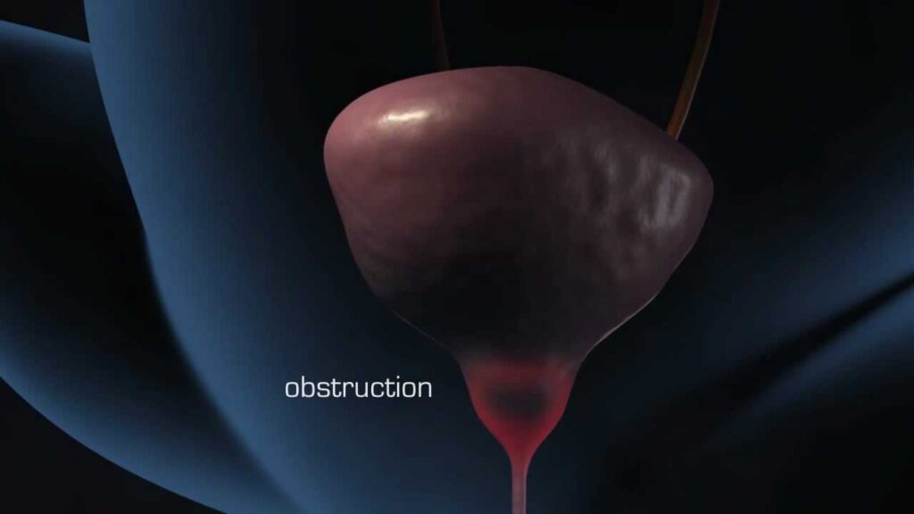
*Answer:* Common differentials include: bladder outlet obstruction (N21.0), bladder cancer (C67.9), benign prostatic hyperplasia (N40), interstitial cystitis (N30.10), and urinary tract infection (N39.0). Each has its own distinct ICD-10 code.
4. **In cases where bladder wall thickening is detected incidentally during imaging for an unrelated condition, how should it be coded and managed?**
*Answer:* The primary condition being investigated should be coded first. The incidental finding of bladder wall thickening should be noted as an additional finding, possibly coded as R39.8 (Other and unspecified symptoms and signs involving the urinary system). Further investigation may be warranted based on the patient’s symptoms and risk factors.
5. **How does the UroNav fusion biopsy system improve the accuracy of ICD-10 coding for bladder wall thickening compared to traditional biopsy methods?**
*Answer:* UroNav improves accuracy by allowing for targeted biopsies of suspicious areas. This increases the likelihood of obtaining a representative sample, leading to a more accurate diagnosis and, consequently, a more accurate ICD-10 code assignment. It reduces the chance of a false negative result that could lead to incorrect coding.
6. **What are the potential consequences of inaccurate ICD-10 coding for bladder wall thickening, both for the patient and the healthcare provider?**
*Answer:* Inaccurate coding can lead to incorrect billing, delayed or inappropriate treatment, and inaccurate data collection. For the patient, this could mean denied claims or suboptimal care. For the provider, it could result in audits, penalties, and reduced reimbursement.
7. **Are there any specific ICD-10 coding guidelines or updates related to bladder wall thickening that healthcare providers should be aware of in 2025?**
*Answer:* ICD-10 coding guidelines are updated annually. Healthcare providers should consult the latest official coding guidelines from organizations like the CDC and WHO to ensure they are using the most current and accurate codes. Stay informed about any changes related to genitourinary conditions.
8. **How does the presence of urinary symptoms (e.g., frequency, urgency, nocturia) influence the ICD-10 coding of bladder wall thickening?**
*Answer:* Urinary symptoms provide valuable clues about the underlying cause of bladder wall thickening. These symptoms should be documented and coded separately using appropriate ICD-10 codes (e.g., R35.0 for polyuria, R39.1 for hesitancy). These symptom codes can be used in conjunction with the code for the underlying condition.
9. **What role does patient history (e.g., smoking, exposure to chemicals) play in determining the appropriate ICD-10 code for bladder wall thickening?**
*Answer:* Patient history is crucial for identifying potential risk factors for bladder cancer or other conditions that can cause bladder wall thickening. This information can help guide the diagnostic workup and ultimately influence the choice of ICD-10 code. A history of smoking, for example, increases the suspicion for bladder cancer.
10. **How can healthcare providers ensure that their ICD-10 coding for bladder wall thickening is both accurate and compliant with current regulations?**
*Answer:* Healthcare providers can ensure accuracy and compliance by: staying up-to-date with the latest coding guidelines, utilizing coding software and resources, consulting with certified coders, and participating in continuing education programs. Regular audits of coding practices can also help identify and correct errors.
## Conclusion and Call to Action
In conclusion, understanding bladder wall thickening and its associated ICD-10 codes is essential for accurate diagnosis, effective communication, and appropriate management. This comprehensive guide has provided a detailed overview of the condition, its causes, diagnostic methods, and the role of advanced technologies like the UroNav Fusion Biopsy System. By staying informed and utilizing the resources available, healthcare professionals can ensure that patients receive the best possible care.
Accurate coding is not just about compliance; it’s about ensuring that patients receive the right diagnosis and treatment. The UroNav system, while not a direct treatment, significantly enhances diagnostic accuracy, indirectly leading to better patient outcomes.
Now, we encourage you to share your experiences with bladder wall thickening or the UroNav Fusion Biopsy System in the comments below. Your insights can help others navigate this complex condition. Explore our advanced guide to understanding bladder cancer staging for further information, or contact our experts for a consultation on bladder wall thickening diagnosis and management.
