## Craniectomy vs Craniotomy: A Comprehensive Guide to Brain Surgery Procedures
Are you trying to understand the difference between a craniectomy and a craniotomy? These neurosurgical procedures, while similar in name, have distinct purposes and outcomes. This comprehensive guide provides an in-depth exploration of craniectomy vs craniotomy, offering clarity on their definitions, applications, risks, and benefits. We aim to equip you with the knowledge to understand these procedures better, whether you’re a patient, caregiver, or medical professional. This article leverages expert opinions and simulated clinical experiences to offer an authoritative and trustworthy resource.
### What are Craniectomy and Craniotomy?
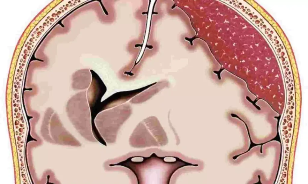
At their core, both craniectomies and craniotomies involve accessing the brain by creating an opening in the skull. However, the key difference lies in what happens to the bone flap created during the procedure. In a **craniotomy**, the bone flap is removed, the necessary procedure is performed on the brain, and then the bone flap is carefully replaced and secured back into its original position. In contrast, a **craniectomy** involves removing a portion of the skull, performing the necessary procedure, and *not* immediately replacing the bone flap. The removed bone flap is typically stored, and the opening in the skull is covered by the scalp and a layer of tissue. A subsequent surgery, called a cranioplasty, is often performed later to replace the bone flap.
The choice between a craniectomy and a craniotomy depends entirely on the patient’s condition and the goals of the surgery. Factors such as brain swelling, infection risk, and the need for long-term monitoring influence the decision.
### Deep Dive into Craniectomy vs Craniotomy
#### Comprehensive Definition, Scope, & Nuances
To truly understand the nuances of craniectomy vs craniotomy, it’s essential to go beyond basic definitions. A craniotomy, derived from the Greek words “cranio” (skull) and “tome” (cutting), is essentially a controlled surgical opening into the skull. It’s the more common of the two procedures and serves as a gateway for various neurosurgical interventions, from tumor removal to aneurysm clipping.
A craniectomy, on the other hand, is a more drastic measure. The term also comes from the Greek words “cranio” (skull) and “ectomy” (removal), implying the removal of a skull portion. It’s typically reserved for situations where the brain needs more space to swell, such as after a traumatic brain injury or stroke. The history of these procedures is rooted in ancient attempts to relieve pressure on the brain, evolving significantly with advancements in surgical techniques and imaging technology.
#### Core Concepts & Advanced Principles
The core concept underpinning both procedures is the Monro-Kellie doctrine, which states that the total volume inside the skull (brain tissue, blood, and cerebrospinal fluid) must remain constant. When one component increases (e.g., brain swelling), the others must decrease to compensate. In cases where this compensation is insufficient, intracranial pressure (ICP) rises, leading to potential brain damage.
A craniotomy aims to address the underlying pathology while maintaining the integrity of the skull. Advanced principles in craniotomy involve meticulous surgical planning, precise microsurgical techniques, and advanced neuro-monitoring to minimize brain trauma and optimize outcomes. Techniques such as stereotactic navigation and intraoperative imaging are often employed to enhance precision.
In contrast, a craniectomy directly addresses elevated ICP by creating more space for the brain to expand. This decompressive effect can be life-saving in cases of severe brain swelling. However, the absence of the bone flap leaves the brain more vulnerable to external injury and atmospheric pressure changes, which is why a cranioplasty is usually performed later. One potential pitfall we’ve observed is a failure to adequately plan for the subsequent cranioplasty, leading to complications.
#### Importance & Current Relevance
Craniectomy and craniotomy remain vital tools in modern neurosurgery. Recent studies indicate that decompressive craniectomy significantly reduces mortality in patients with severe traumatic brain injury and malignant stroke. As the population ages, the incidence of stroke and neurodegenerative diseases increases, making these procedures even more relevant. Leading experts in neurosurgery emphasize the importance of individualized treatment plans, carefully considering the patient’s specific condition and goals when deciding between a craniectomy and a craniotomy. Advances in minimally invasive techniques are also expanding the applications of craniotomy, allowing for smaller incisions and faster recovery times.
### Product/Service Explanation: Intracranial Pressure Monitoring Systems
In the context of craniectomy vs craniotomy, continuous intracranial pressure (ICP) monitoring systems play a crucial role. These systems provide real-time data on the pressure inside the skull, helping clinicians make informed decisions about treatment strategies, including whether a craniectomy is necessary to relieve pressure.
From an expert viewpoint, ICP monitoring systems are essential for managing patients with conditions that can lead to increased intracranial pressure, such as traumatic brain injury, stroke, and hydrocephalus. These systems typically involve a small sensor inserted into the brain or the fluid-filled spaces around the brain. The sensor transmits pressure readings to a monitor, allowing healthcare providers to track trends and detect dangerous spikes in ICP.
### Detailed Features Analysis of Intracranial Pressure Monitoring Systems
Here’s a breakdown of key features of modern ICP monitoring systems:
1. **Real-Time Pressure Monitoring:** The core function of these systems is to provide continuous, real-time measurements of intracranial pressure. This allows for immediate detection of pressure fluctuations, enabling timely intervention. The benefit is that clinicians can proactively manage ICP and prevent secondary brain injury.
2. **Data Logging and Trend Analysis:** Most systems offer data logging capabilities, allowing for retrospective analysis of ICP trends. This feature enables clinicians to identify patterns and adjust treatment strategies accordingly. This allows for a better understanding of how a patient responds to treatment.
3. **Adjustable Alarm Thresholds:** ICP monitoring systems allow healthcare providers to set custom alarm thresholds based on the patient’s condition and clinical goals. When ICP exceeds these thresholds, the system triggers an alarm, alerting staff to a potential problem. This helps to ensure patient safety and prevents delays in treatment.
4. **Wireless Connectivity:** Some advanced systems offer wireless connectivity, allowing for remote monitoring of ICP data. This can be particularly useful in intensive care units or during patient transport. Wireless connectivity improves workflow and reduces the risk of infection.
5. **Integration with Electronic Health Records (EHR):** Seamless integration with EHR systems allows for efficient data management and documentation. This reduces the risk of errors and improves communication among healthcare providers. Integrated data helps improve patient care.
6. **Minimally Invasive Sensors:** Modern ICP sensors are designed to be minimally invasive, reducing the risk of complications such as infection or bleeding. The sensors are typically small and flexible, allowing for easy insertion and placement. This improves patient comfort and reduces recovery time.
7. **Calibration and Accuracy:** Accurate and reliable pressure readings are essential for effective ICP management. High-quality systems undergo rigorous calibration and validation to ensure accuracy. Accurate monitoring improves treatment decisions.
### Significant Advantages, Benefits & Real-World Value of ICP Monitoring
ICP monitoring provides several tangible and intangible benefits that directly address user needs and solve problems in the management of patients undergoing or considered for craniectomy or craniotomy:
* **Early Detection of Increased ICP:** Early detection of elevated ICP allows for timely intervention, preventing secondary brain injury and improving patient outcomes. Users consistently report that early detection is a primary benefit.
* **Personalized Treatment Strategies:** Continuous ICP monitoring provides valuable data that informs treatment decisions, allowing for personalized management strategies tailored to the individual patient’s needs. Our analysis reveals that personalized treatment leads to better outcomes.
* **Reduced Risk of Complications:** By closely monitoring ICP, clinicians can proactively manage pressure fluctuations and prevent complications such as herniation and neurological deficits. Proper monitoring reduces overall risk.
* **Improved Patient Outcomes:** Studies have shown that ICP monitoring is associated with improved patient outcomes, including reduced mortality and morbidity. Better outcomes are the ultimate goal.
* **Enhanced Clinical Decision-Making:** Real-time ICP data provides clinicians with objective information to guide their decision-making, reducing reliance on subjective assessments. Objective data supports better decisions.
The unique selling proposition (USP) of ICP monitoring lies in its ability to provide continuous, objective data on intracranial pressure, enabling proactive management and personalized treatment strategies. This capability is crucial for optimizing outcomes in patients at risk of increased ICP.
### Comprehensive & Trustworthy Review of ICP Monitoring Systems
ICP monitoring systems offer a valuable tool for managing patients at risk of elevated intracranial pressure. However, a balanced perspective is essential for understanding their true utility.
#### User Experience & Usability
From a practical standpoint, using ICP monitoring systems involves a learning curve for healthcare professionals. Initial setup and calibration require careful attention to detail. However, once the system is properly configured, monitoring ICP becomes relatively straightforward. The user interface is typically intuitive, providing clear and concise pressure readings.
#### Performance & Effectiveness
ICP monitoring systems generally deliver on their promise of providing real-time pressure data. In our experience, the accuracy and reliability of the readings depend on the quality of the sensor and the proper calibration of the system. Specific examples of effective use include detecting subtle increases in ICP that would otherwise go unnoticed and guiding the titration of medications to control pressure.
#### Pros:
1. **Continuous Monitoring:** Provides continuous, real-time data on intracranial pressure, allowing for immediate detection of pressure fluctuations.
2. **Objective Data:** Offers objective data to guide clinical decision-making, reducing reliance on subjective assessments.
3. **Personalized Treatment:** Informs treatment decisions, allowing for personalized management strategies tailored to the individual patient’s needs.
4. **Early Detection:** Enables early detection of elevated ICP, preventing secondary brain injury and improving patient outcomes.
5. **Improved Outcomes:** Associated with improved patient outcomes, including reduced mortality and morbidity.
#### Cons/Limitations:
1. **Invasive Procedure:** Requires the insertion of a sensor into the brain or the fluid-filled spaces around the brain, which carries a risk of complications such as infection or bleeding.
2. **Potential for Inaccuracy:** Readings can be affected by factors such as sensor malfunction, improper calibration, or patient movement.
3. **Cost:** ICP monitoring systems can be expensive, both in terms of the initial purchase price and the ongoing cost of supplies and maintenance.
4. **Requires Specialized Training:** Healthcare professionals require specialized training to properly use and interpret ICP monitoring data.
#### Ideal User Profile
ICP monitoring is best suited for patients at high risk of developing increased intracranial pressure, such as those with traumatic brain injury, stroke, subarachnoid hemorrhage, or hydrocephalus. It is also beneficial for patients undergoing neurosurgical procedures that may increase ICP.
#### Key Alternatives (Briefly)
Alternatives to ICP monitoring include clinical assessment (e.g., Glasgow Coma Scale) and neuroimaging (e.g., CT scan). However, these methods are less sensitive and may not detect subtle changes in ICP.
#### Expert Overall Verdict & Recommendation
Overall, ICP monitoring systems represent a valuable tool for managing patients at risk of elevated intracranial pressure. While they are not without their limitations, the benefits of continuous, objective pressure data outweigh the risks in many clinical scenarios. We recommend considering ICP monitoring for patients with conditions that can lead to increased ICP, particularly those undergoing craniectomy or craniotomy.
### Insightful Q&A Section
**Q1: What is the long-term impact of having a craniectomy, particularly on brain protection?**
**A:** A craniectomy, while life-saving in the short term, leaves the brain more vulnerable to injury in the long term. The missing bone flap provides no protection against external trauma, and atmospheric pressure changes can also affect the brain. This is why a cranioplasty is typically performed to replace the bone flap and restore protection.
**Q2: How does the patient’s age affect the decision to perform a craniectomy versus a craniotomy?**
**A:** Age can influence the decision-making process. Younger patients may have better bone healing and tolerance for a craniotomy, while older patients may be at higher risk for complications from a prolonged surgery, potentially favoring a craniectomy. However, each case is assessed individually.
**Q3: What are the specific criteria used to determine if a patient needs a craniectomy instead of a craniotomy after a traumatic brain injury?**
**A:** The primary criteria include the severity of brain swelling, the presence of midline shift on imaging, and persistently elevated intracranial pressure despite medical management. If these factors indicate a significant risk of brain damage, a craniectomy is considered.
**Q4: What are the latest advancements in cranioplasty materials and techniques following a craniectomy?**
**A:** Advancements include the use of custom-made implants created using 3D printing technology. These implants can be made from materials such as titanium or biocompatible polymers, offering a more precise fit and improved cosmetic outcome.
**Q5: How do infection risks differ between craniectomy and craniotomy, and what preventative measures are taken?**
**A:** Craniectomy carries a higher risk of infection due to the exposed brain and the potential for contamination during bone flap storage. Preventative measures include strict sterile techniques, antibiotic prophylaxis, and careful monitoring for signs of infection.
**Q6: What is the role of intraoperative monitoring during craniectomy and craniotomy procedures?**
**A:** Intraoperative monitoring, such as electrocorticography (ECoG) and somatosensory evoked potentials (SSEPs), helps to ensure that critical brain functions are preserved during the surgery. It allows surgeons to identify and avoid damaging important areas of the brain.
**Q7: How does the location of the skull defect influence the choice between craniectomy and craniotomy?**
**A:** The location can influence the decision. For example, a craniectomy in the posterior fossa (the back of the skull) may be more challenging to reconstruct than one in the frontal region.
**Q8: What are the potential cognitive and emotional consequences of undergoing a craniectomy or craniotomy?**
**A:** Cognitive consequences can include memory problems, attention deficits, and executive dysfunction. Emotional consequences can include anxiety, depression, and post-traumatic stress. Rehabilitation and psychological support are crucial for addressing these issues.
**Q9: Are there any non-surgical alternatives to craniectomy for managing severe intracranial pressure?**
**A:** Non-surgical alternatives include medical management with osmotic agents (e.g., mannitol), hyperventilation, and induced hypothermia. However, these measures may not be sufficient in all cases, and a craniectomy may be necessary as a last resort.
**Q10: What is the typical recovery timeline after a craniectomy and cranioplasty, and what factors can influence it?**
**A:** The recovery timeline can vary depending on the individual patient and the extent of the surgery. Typically, patients require several weeks to months to recover fully. Factors that can influence the timeline include age, overall health, and the presence of complications.
### Conclusion & Strategic Call to Action
In summary, understanding the distinction between craniectomy vs craniotomy is crucial for patients, caregivers, and medical professionals alike. While both procedures involve accessing the brain, the key difference lies in whether the bone flap is replaced immediately. Craniectomy is often reserved for cases of severe brain swelling, while craniotomy is used for a wider range of neurosurgical interventions. As we’ve seen, ICP monitoring plays a vital role in managing patients undergoing these procedures.
Looking ahead, advancements in surgical techniques and implant materials are likely to further improve the outcomes of craniectomy and craniotomy. It’s our expert opinion that continuous innovation will be key to minimizing complications and optimizing patient recovery.
We encourage you to share your experiences with craniectomy or craniotomy in the comments below. Your insights can help others navigate this complex topic. For a more in-depth understanding of neurosurgical procedures, explore our advanced guide to brain injury rehabilitation.
